Labelled Diagram Of Plant Cell Under Electron Microscope / Topic Labeling Animal And Plant Cells Under The - Here's a photo of a plant cell under an electron microscope.
Labelled Diagram Of Plant Cell Under Electron Microscope / Topic Labeling Animal And Plant Cells Under The - Here's a photo of a plant cell under an electron microscope.. Draw a fully labelled diagram of a plant cell as seen. Cautionary labels are given for products or containers containing hazardous material. The detail that can be seen, or resolution, is also important. You can specify conditions of storing and accessing cookies in your browser. Interpretation of electron micrographs to identify organelles and deduce the function of specialised cells.
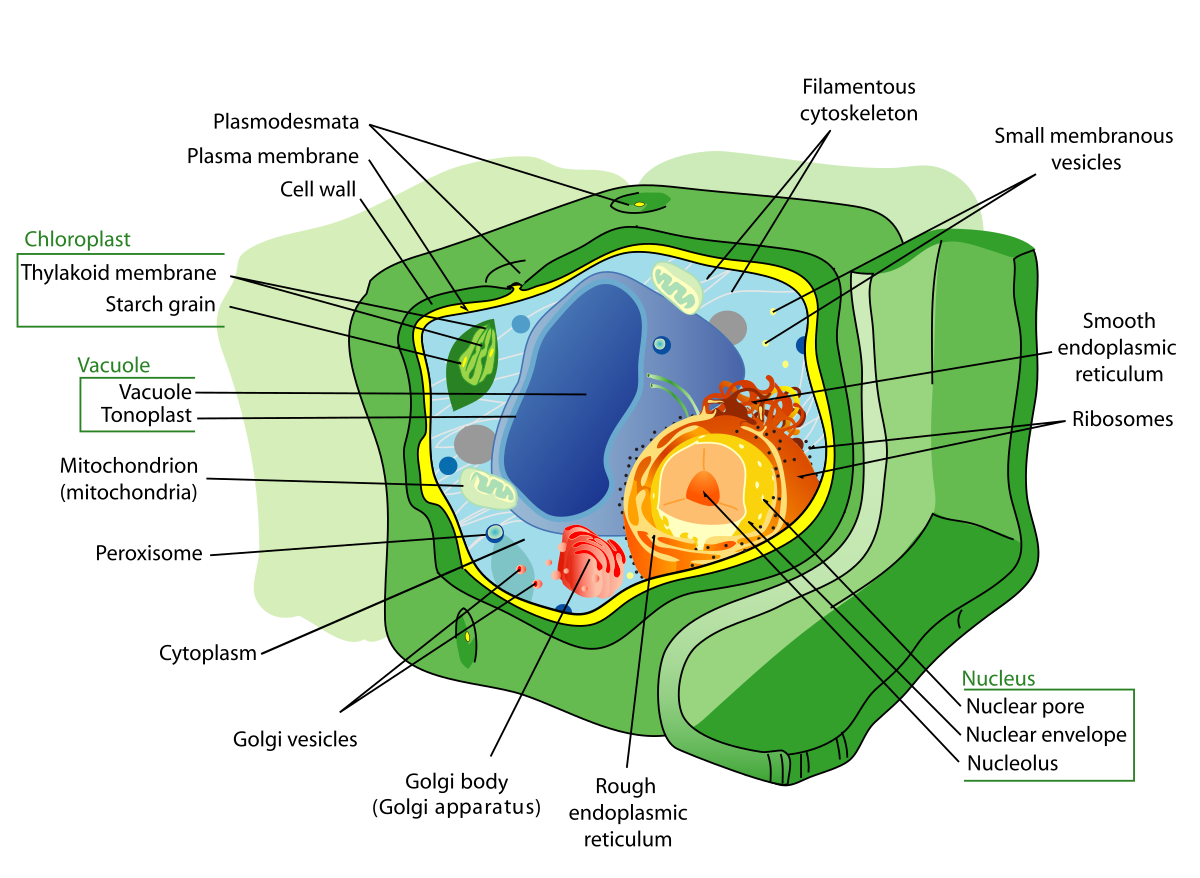
The term 'cell' was coined to describe the small walled units that were observed in the sections of bottle cork under simple microscope. Interpretation of electron micrographs to identify organelles and deduce the function of specialised cells. Plant cells contain many organelles such as ribosomes, the nucleus, the plasma membrane, the cell wall, mitochondria, and chloroplasts. Ishita observed a slide of eukaryotic cell under electron microscope. (i) name the parts labelled as 1 to 10.
Plant Cell Wikipedia from upload.wikimedia.org Besides identification which is a major purpose of labels. You can specify conditions of storing and accessing cookies in your browser. They are cells that have a distinct nucleus and other cellular organelles under the microscope, it shows many different parts. Robert hooke in 1665 first discovered plant cell. Each part, known as an organelle, works together to keep the cell functional. The diagram shows part of a cell surface membrane. Plant cell structure under microscope : The typical characteristics that define the plant cell include cellulose, hemicellulose and pectin, plastids which play a major role in photosynthesis and storage of starch, large vacuoles responsible for regulating the cell turgor pressure.
The magnification of a microscope is not the only factor that is important when viewing cells.
These are easily observed under the electron microscopy has revealed that the nuclear envelope, which consists of two parallel. The electron microscope sends a stream of electrons through a vacuum. (i) name the parts labelled as 1 to 10. Does anyone have a decent labelled diagram of a plant cell under an electron microscope? What was once unseeable can now be seen, touched, and eaten!cut. Plant cell science diagram clipart set includes: Draw a fully labelled diagram of a plant cell as seen. The diagram is taken from an electron micrograph of a cell which. Labels are a means of identifying a product or container through a piece of fabric, paper, metal or plastic film onto which information about them is printed. Here's a photo of a plant cell under an electron microscope. Each part, known as an organelle, works together to keep the cell functional. Most animals and plants are composed of many millions of cells, but some organisms such as bacteria and protoctistans consist of single cells. When you look at animal or plant cells under the electron microscope, you can see a lot more detail.
The diagram is very clear, and labeled the diagram is very clear, and labeled; 719 x 539 jpeg 58 кб. Examining a diagram of the plant cell will help make the differences clearer. The structure of a cell can be studied under a light or electron microscope. Plant, animal and bacterial cells have smaller components each with a specific function.
Draw A Neat Diagram Of Plant Cell And Label Any Three Parts Which Differentiate It From Animal Cell Studyrankersonline from www.studyrankersonline.com Which structures would be clearly visible at a magnification of 400? Besides identification which is a major purpose of labels. When you look at animal or plant cells under the electron microscope, you can see a lot more detail. In the given figure of an animal cell as observed under an electron microscope. The plasma membrane, also a cell wall in plant cells. The diagram is very clear, and labeled the diagram is very clear, and labeled; Examining a diagram of the plant cell will help make the differences clearer. As you guessed, it is photosynthesis.
The detail that can be seen, or resolution, is also important.
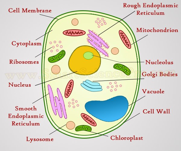
Plant, animal and bacterial cells have smaller components each with a specific function. Here's a diagram of a plant cell: As you can see in the above labeled plant cell diagram under light microscope, there are 13 plant leaves are capable of doing something highly important to earth. Transmission electron microscopy is a proven technique in the field of cell biology and a very useful tool in biomedical research. Draw a fully labelled diagram of a plant cell as seen. The diagram is very clear, and labeled the diagram is very clear, and labeled; While organelles have identifying structures, specific shapes may vary. Most animals and plants are composed of many millions of cells, but some organisms such as bacteria and protoctistans consist of single cells. In the given figure of an animal cell as observed under an electron microscope. 1135 x 1135 jpeg 179 кб. Explanation:i know how to draw diagram. Robert hooke in 1665 first discovered plant cell. Electron microscopic studies of eukaryotic cells reveal the presence of a network or reticulum of tiny tubular plastids are found in all plant cells and in euglenoides.
Cautionary labels are given for products or containers containing hazardous material. Each part, known as an organelle, works together to keep the cell functional. Plant cells contain many organelles such as ribosomes, the nucleus, the plasma membrane, the cell wall, mitochondria, and chloroplasts. Major differences between a plant cell and on animal cell are (i) presence of draw a neat diagram of plant cell and label any three parts which differentiate it from animal cell. The diagram is very clear, and labeled the diagram is very clear, and labeled;
Structure Of Animal Cell And Plant Cell Under Microscope Diagrams from www.smartsciencepro.com Draw a fully labelled diagram of a plant cell as seen. This is a single cheek cell that has been stained, as seen under a light microscope. A micrograph is a photo or digital image taken through a microscope to show a magnified image of a specimen. Plant, animal and bacterial cells have smaller components each with a specific function. In the given figure of an animal cell as observed under an electron microscope. Electron microscopic studies of eukaryotic cells reveal the presence of a network or reticulum of tiny tubular plastids are found in all plant cells and in euglenoides. They are cells that have a distinct nucleus and other cellular organelles under the microscope, it shows many different parts. Plant cells are the basic unit and building blocks of life in organisms of the kingdom plantae.
They are cells that have a distinct nucleus and other cellular organelles under the microscope, it shows many different parts.
They are cells that have a distinct nucleus and other cellular organelles under the microscope, it shows many different parts. Besides identification which is a major purpose of labels. Here's a photo of a plant cell under an electron microscope. Examining a diagram of the plant cell will help make the differences clearer. The typical characteristics that define the plant cell include cellulose, hemicellulose and pectin, plastids which play a major role in photosynthesis and storage of starch, large vacuoles responsible for regulating the cell turgor pressure. Transmission electron microscopy is a proven technique in the field of cell biology and a very useful tool in biomedical research. Which structures would be clearly visible at a magnification of 400? When you look at animal or plant cells under the electron microscope, you can see a lot more detail. Major differences between a plant cell and on animal cell are (i) presence of draw a neat diagram of plant cell and label any three parts which differentiate it from animal cell. In the given figure of an animal cell as observed under an electron microscope. The diagram shows part of a cell surface membrane. A micrograph is a photo or digital image taken through a microscope to show a magnified image of a specimen. As you can see in the above labeled plant cell diagram under light microscope, there are 13 plant leaves are capable of doing something highly important to earth.
The detail that can be seen, or resolution, is also important plant cell under electron microscope labelled. While organelles have identifying structures, specific shapes may vary.


0 Comments